Stereotactic biopsy is a procedure that enables a neurosurgeon to extract a small piece of tissue with a thin needle and use it for diagnosis by testing it under a microscope. It is very useful for diagnosing brain lesions. Whenever initial scans and tests reveal uncertain results, a stereotactic biopsy surgeryis necessary to narrow down the issue and then take a decision about the treatment. It allows a neurosurgeon to map the brain into three dimensional coordinate system and then select the exact location for performing the biopsy.
The number of patients affected by primary brain tumors and meta static tumors are increasing and neurosurgeons need to have enough ways to diagnose and detect them accurately to define a treatment plan. Initially, Magnetic Resonance Imaging is used to provide information about the exact location of the tumor. The size and it’s relationship to the surrounding structures and cells can also be understood. Magnetic resonance spectroscopy is then performed to understand the chemical composition of the tumor. Profusion and diffusion provides information about the water and blood flow through the tumor.
A more definitive way of diagnosing the exact nature of a tumor is by biopsy – or by extracting a piece of the tissue and examining it under a microscope and performing certain other tests outside.
Technique Used
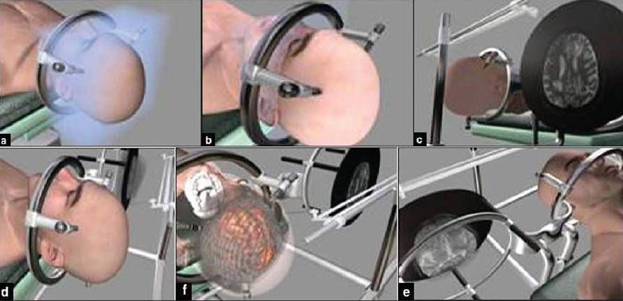
In this procedure, the patient is awake and the head is secured. The fiducials on the scalp are registers by cameras and can be seen on a computerized navigation system in the operating room. In order to make an incision, a small part of the head is shaved to make a marking where the incision will be made. The area is cleaned and is draped to maintain it in a sterile way. A small opening in the brain is made by passing through the skull. The stereotactic needle that has a long, blunt and soft nosed tip is inserted and guided through the opening with the help of the neuro navigation system. In this way samples of the tumor are obtained.
These samples are examined in real time by pathologists. Also, samples are retrieved for detailed examination later on. It takes a pathologist 3-4 days to complete all the tests and derive a conclusion from the samples. In this way they get more accurate readings and can help in deciding the further course of treatment.
After this, the incision is carefully closed. Clean and dry dressing is applied. This takes two to three days to recover and remove it.
Hospital stay and other factors
The hospital stay for this procedure is generally short and requires just one night. You will be free to go home after that. The sutures are generally removed after fifteen days.
This procedure is most commonly used to treat brain ailments such as tumors, infections and neurodegenerative diseases.
This procedure is very effective in deciding whether a patient is a good candidate for surgery or alternative medication can help instead.
After this procedure, make sure that you keep your appointment, so that if any infections or risk factors are detected, they can be detected and treated in the early stages!